
Figure 4. Administration of bretazenil into the PBN prevents starvation in mice after ablation of AgRP neurons.
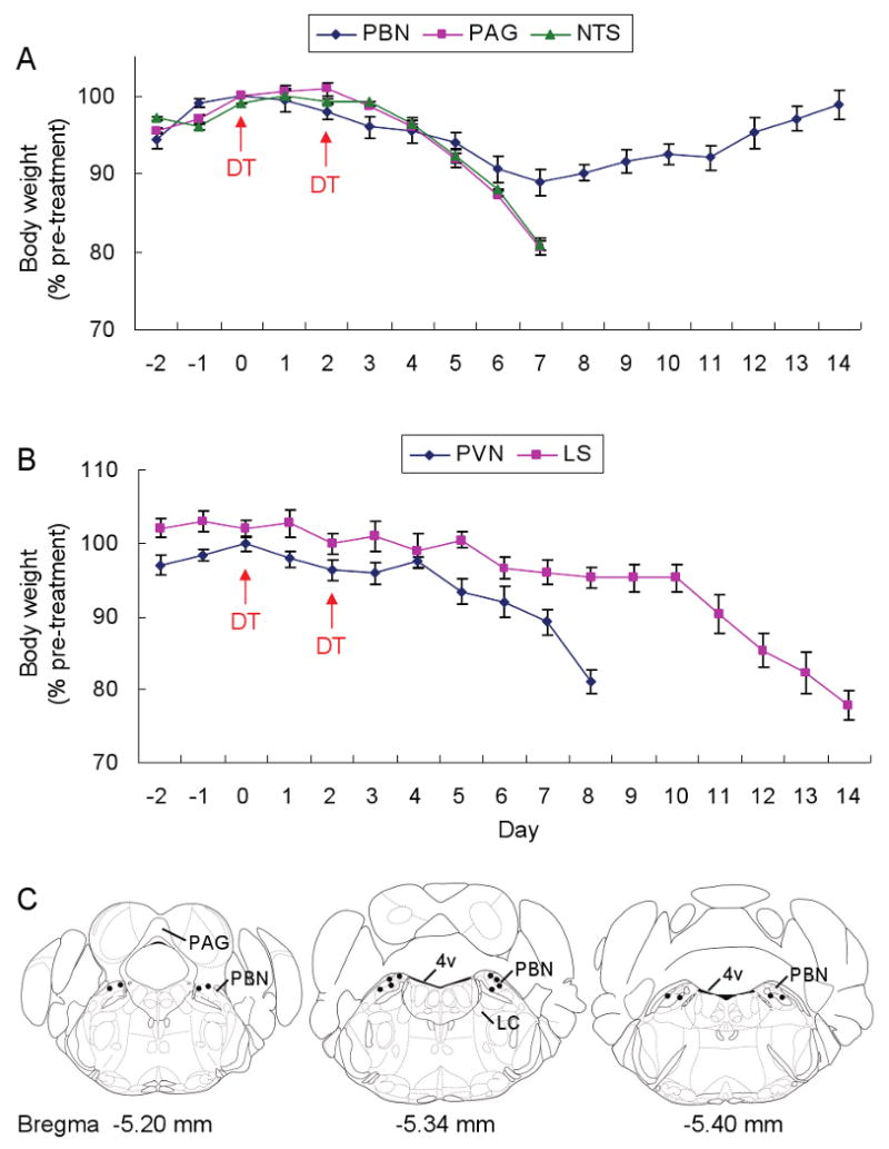
(A) Percentage of initial body weight of DT-treated, AgrpDTR/+ mice after chronic administration of bretazenil in 3 post-synaptic areas of AgRP neurons in the hindbrain (PBN, PAG, and NTS).
(B) Percentage of initial body weight of DT-treateted, AgrpDTR/+ mice after chronic administration of bretazenil in 2 forebrain regions (PVN and LS) with projections from AgRP neurons.
N = 6 – 8 per each group. Error bars represent the SEM.
(C) Coronal illustrations of mouse hindbrain regions; the dots represent effective infusion sites in the PBN area. LC, locus ceruleus; 4v, 4th ventricle.