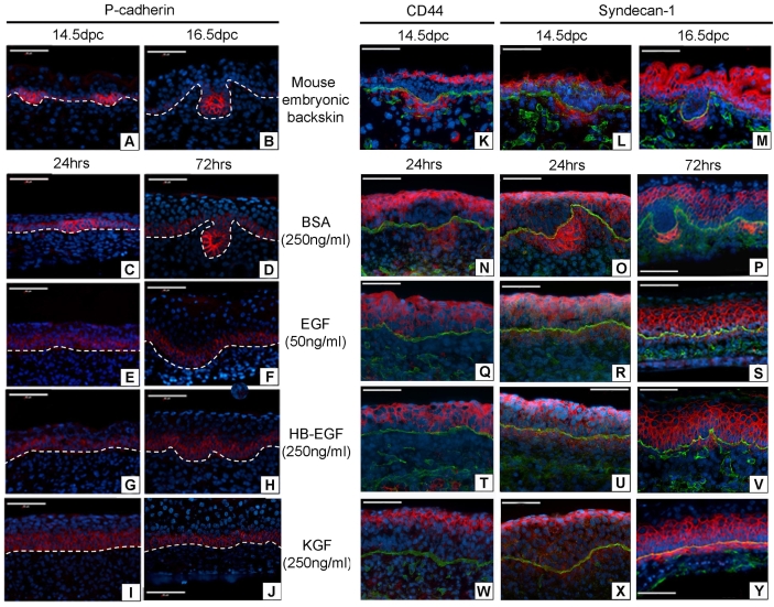
Fig. 4.
P-cadherin, CD44 and syndecan 1 confirm the absence of placodes and dermal condensations in ligand-treated skin. (A-Y) P-cadherin, CD44 and syndecan 1 expression (red) in ligand-treated mouse back skin. P-cadherin is upregulated at sites of placode formation during HF morphogenesis (A,B). CD44 and syndecan 1 are expressed only in the DC within the dermal compartment of embryonic skin during HF morphogenesis (K-M). Controls show a distribution of P-cadherin, CD44 and syndecan 1 comparable with in vivo embryonic skin (C,D,N-P). Skin cultured with EGFR ligands EGF (50 ng/ml) or HBEGF (250 ng/ml) displayed no localised upregulated P-cadherin expression (E-H) and no differential dermal expression of CD44 or syndecan 1 (Q-V). Skin cultured with KGF (250 ng/ml) displayed no localised upregulated P-cadherin expression (I,J) or CD44 (W). KGF treatment resulted in a continuous layer of syndecan 1 expression in the dermal cells subjacent to the epidermis (X,Y). P-cadherin, CD44 and syndecan 1 (red), laminin (green), nuclear DAPI (blue). Scale bars: 60 μm.

