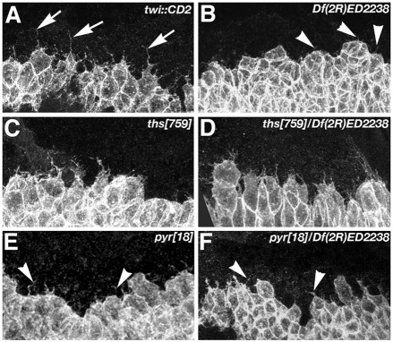
Fig. 3.
Morphology of dorsal edge cells during mesoderm migration in FGF8-like ligand mutants. Cell shape at the dorsal edge of the mesoderm during dorsolateral migration visualised using twi::CD2. In wild type (twi::CD2), dorsal edge cells form thin and long protrusions (A, arrows). Only short, filopodial protrusions (arrowheads) are present in embryos lacking pyr gene function (B,E,F). In ths759 homozygous (C) or hemizygous (D) embryos, long protrusions are present similar to those in the wild type. Note that cells at the dorsal edge in E,F fail to extend along the dorsal ventral axis (dorsal is up and ventral is down).

