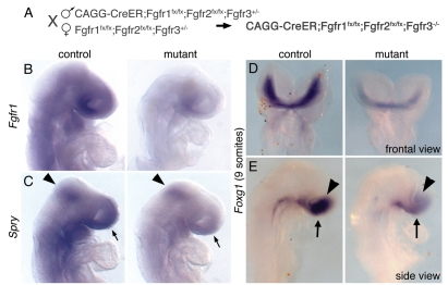
Fig. 6.
Reduced FGF signaling prior to telencephalon development leads to reduced Foxg1 expression. (A) To disrupt FGF signaling earlier than the start of telencephalon development, a CAGG-CreER allele was used to recombine floxed alleles of Fgfr1 and Fgfr2 in an Fgfr3-null background using tamoxifen administration at E6.75 and E7.75. (B) Expression of Fgfr1 at the 11- to 12-somite stage, which is found ubiquitously in the control (left), is reduced but not abolished in the mutant (right). (C) Similarly, at the same stage, Spry1 expression is reduced, but not abolished, in the isthmus (arrowhead), anterior neurectoderm (arrow) and facial tissue of the mutant. (D,E) Slightly earlier (E8.5, 9-somite stage), when Foxg1 has just started to be expressed in the control embryo (left), it is already significantly reduced in the anterior neuroepithelium (arrowhead) and to a lesser extent in the anterior neural ridge (arrow) of the mutant (right). D, frontal view; E, side view.

