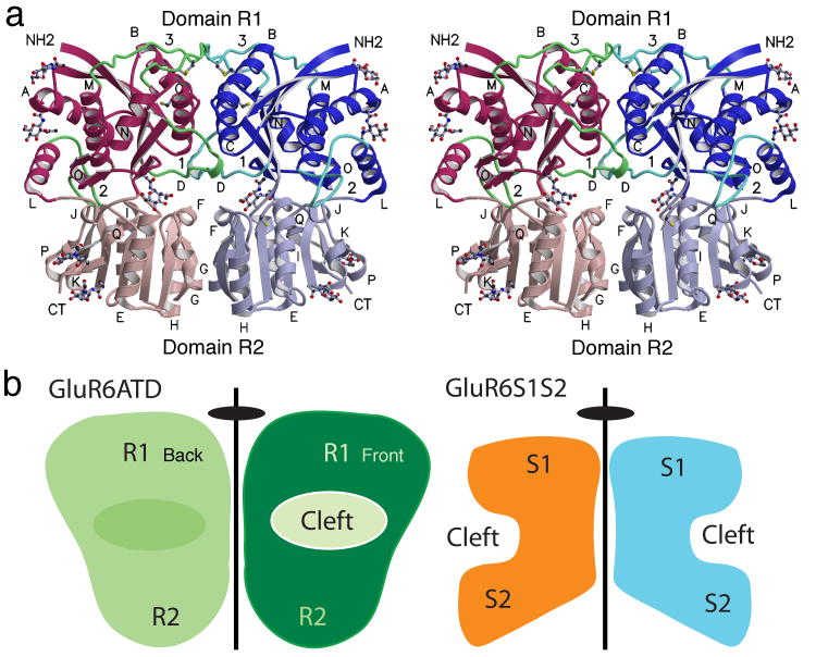
Figure 3.
The GluR6 amino terminal domain crystallizes as a dimer. (a) Stereoview of the dimer assembly with domains R1 and R2 from each subunit shaded in dark and light red and blue respectively; loops 1-3 are colored green; N-linked NAG molecules and Cys side chains are drawn in ball and stick representation; α-helices are labeled for each subunit. The cleft between domains 1 and 2 for the left subunit is on the rear plane of the dimer, while for the right subunit the cleft faces the viewer resulting in a back to front arrangement with respect to the dimer axis of symmetry. (b) Cartoons showing the location of the clefts between domains 1 and 2 relative to the dimer 2-fold axis of symmetry in the GluR6 ATD dimer (left) and the GluR2 S1S2 ligand binding domain dimer (right). The view for the ATD cartoon matches that in (a), for which the interdomain cleft for the left hand subunit is not visible.

