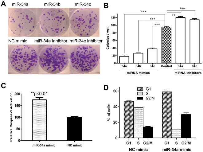
Figure 3. Restoration of miR-34 inhibits the clonogenic growth of MiaPaCa2 cells, whereas inhibition of miR-34 promotes cell growth.
MiaPaCa2 cells were transfected with miR-34 mimics or inhibitors, 24 hr later the cells were seeded in 6-well plates (200 cells/well, in triplicates). After 12–14 days incubation, the plates were gently washed with PBS and stained with 0.1% crystal violet. A, representative pictures of the colonies. B, Colonies with over 50 cells were counted. C, Restoration of miR-34 leads to caspase-3 activation. Caspase-3 activation assay was carried out as described in in Materials and Methods . Fold increase of fluorescence signal was calculated by dividing the normalized signal in each treated sample with that in the untreated control. **P<0.01, ***P<0.001, Student's t-test, n = 3. D, Cell cycle distribution of MiaPaCa2 cells transfected with miR-34 mimics. Cell cycle analysis was performed 1 day after transfection. Cells were stained with propidium iodide after ethanol fixation and analyzed by flow cytometry.

