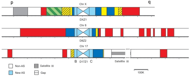
Figure 2. Alpha satellite layers in human chromosomes 8, 17, and X.
Each colored domain represents an AS array composed of monomers that belong to the same branch on phylogenetic trees shown in Figure 1. Chromosome domains and the arches marking different branches are in the same colors in Figures 1 and 2. Colored layers are partially symmetrical around the centromere on one chromosome and partially shared between different chromosomes. The p and q arms of the chromosomes are indicated. The diagonally crossed white and light blue central boxes represent the new AS HOR domains, which form current centromeres. They are shown not to scale. For chromosome 17, we show the presumed organization of the HOR domain. The central D17Z1 16-mer HOR array is flanked by two homogenous 14-mer HOR arrays, D17Z1-B on the p arm [24], and a distinct one termed D17Z1-C on the q arm (see Text S1 for details).

