
Figure 5.
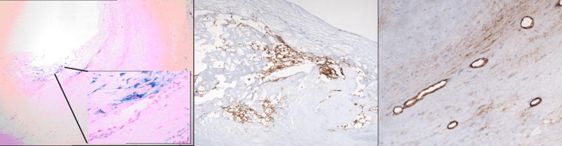
Plaque Histopathology. Left panel: Histopathological section with Prussian Blue staining at low and high (inset) magnifications demonstrate iron deposits in plaque. Middle panel: Staining for glycophorin A shows evidence of red blood cell membrane fragments within plaque. Right panel: Staining for Factor VIII demonstrates neovascularization in the plaque neointima.