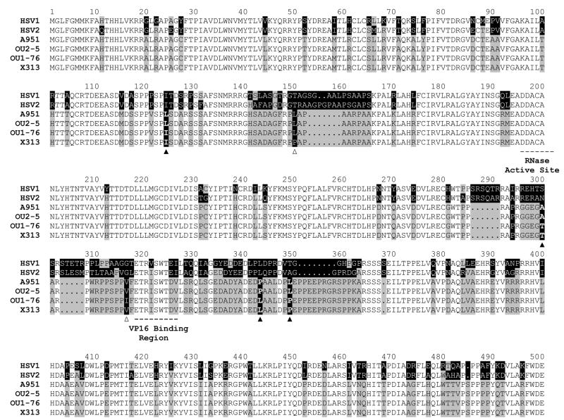
Figure 4. Location of variable amino acid residues in the HVP2 UL41 (vhs) polypeptide.
AA sequences of four HVP2 strains (HVP2ap A951 & OU2-5 and HVP2nv OU1-76 & X313) were aligned to identify residues that varied among isolates. The sequences for HSV1 and HSV2 are included for reference. AA residues conserved in all sequences are indicated by lack of shading. Residues that vary between HSV and HVP2 are indicated by gray/black shading. The four potential HVP2 subtype-specific substitutions are indicated by a solid triangle below the sequences and the two isolate-specific substitutions are indicated by an open triangle below them.

