
Figure 5. Characterization of UL41 mutants.
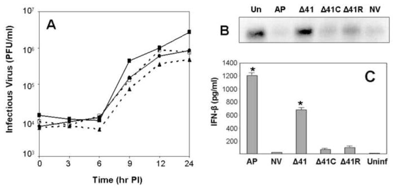
One-step growth curves performed in Vero cells are shown in A. Viruses included the parental HVP2nv (■——-■), HVP2nv Δ41 (▲………▲), HVP2nv Δ41C (□----□) and HVP2nv Δ41R (●——-●). Northern blot of β-actin mRNA in infected cells is shown in B. Total RNA was isolated from cells at 4 hrs PI, 15 μg loaded in each lane, and northern blotting performed using the Vero cell β-actin gene as probe. IFN-β production in PMDF cultures is shown in C. Cultures were infected with the indicated viruses at an MOI = 1 PFU/cell and the medium assayed for IFN-β at 24 hrs PI. IFN-β levels that were significantly different (p = >0.05) from the level induced by the parental HVP2nv virus are indicated by an asterisk above the standard error bar.