
Figure 3. Regulatory T cells depletion combined with interleukin-2 (IL-2) permits tumor localization of vesicular stomatitis virus (VSV).
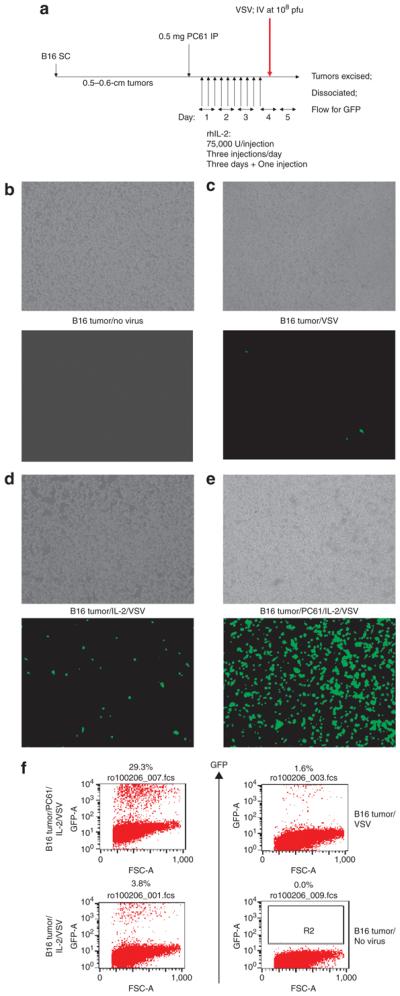
(a) C57Bl/6 mice (three/group) were seeded with subcutaneous (SC) B16 tumors. After 9 days, the mice received an intraperitoneal (IP) injection of PC61 or control immunoglobulin G (IgG). After a further 24hours, the mice were injected IP with phosphate-buffered saline (PBS) or with recombinant human (75,000U/injection three times a day for 3 days). On the fourth day, a single further injection of IL-2 was given. Two hours after this last injection of IL-2/PBS, the mice received an intravenous (IV) injection of 108 plaque forming units (pfu) of VSV-GFP. (b–e) Thirty-six hours after in vivo virus delivery, the tumors were explanted and dissociated in vitro. The cells were left to adhere to the culture dish for 6–12 hours and then washed three times gently in PBS to remove nonadherent cells. These cultures were examined visually using phase microscopy (upper panels) or fluorescence for GFP expression (lower panels). The treatments received by the mice from which the tumors were explanted are shown between the panels. This protocol reproducibly generated cultures which were >98% positive for gp100, a B16-specific melanoma/melanocyte-associated antigen. This shows that GFP positivity is derived almost exclusively from recovered tumor cells rather than from infiltrating (nonadherent) immune cells [such as natural killer (NK) cells]. (f) Samples of the explanted B16 tumors obtained from a–e above were analyzed using flow cytometry for GFP. The results from a–f are representative of three separate experiments. GFP, green fluorescent protein. FSC, forward scatter.