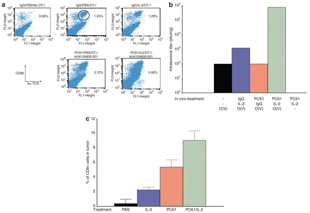
Figure 2. Treg depletion+IL-2 increases T-cell persistence and virus delivery.
(a) B16ova tumors were established subcutaneously in C57Bl/6 mice. Ten days later, following the regimen described in Figure 1a, mice received either a control IgG or anti-CD25 antibody PC-61; 1 day later this was followed by 10 i.p. injections of phosphate-buffered saline (PBS) or of rhIL-2 at a dose of 75,000 U/injection; 24 hours after the last injection of PBS or IL-2, mice received an intravenous injection of either PBS or of 106 OT-I cells. Tumors were harvested 48 hours later, dissociated and analyzed by flow cytometry for OT-I T cells, expressed as the percentage of cells from the tumors which labeled double positive for both CD8 and the Vα2 T-cell receptor expressed by OT-I transgenic mice. Data shown are from individual mice from groups of three mice/treatment and are representative of two separate experiments. (b) The experiment of a was repeated with mice treated with PC-61 and/or IL-2 as shown; 24 hours after the last injection of IL-2/PBS, groups received an intravenous injection of 106 OT-I loaded with VSV at an MOI of 1 (O(V)) or PBS (−). Forty-eight hours later, mice were euthanized and viral titers recovered from the freeze thaw/lysates of the tumors. Viral titers are the mean of two mice per group. Data shown are representative of two separate experiments. (c) The experiment of a was repeated with mice (3/group) treated with PBS, IL-2 alone, PC-61 alone, or PC-61+IL-2. Seventy-two hours after the last injection of IL-2/PBS, tumors were harvested, dissociated, and analyzed by flow cytometry for CD3+, CD8+ T cells. Values are expressed as the percentage of cells from the tumors which labeled double positive for both CD8 and CD3. Data are representative of two separate experiments.

