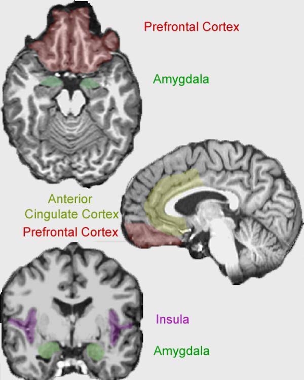
Figure 1.
TOP: A low transverse MRI image of the prefrontal cortex (in red) and the amygdala (in green). MIDDLE: A mid-sagittal MRI image of the anterior cingulate cortex (in yellow) and the prefrontal cortex (in red). BOTTOM: A mid-coronal MRI image of the insula (in purple) and the amygdala (in green). It should be noted that the neuroanatomical distinction between the ventromedial prefrontal cortex and the orbitofrontal cortex is not well-delineated. Though the orbitofrontal cortex may be closer to the eyes than the ventromedial prefrontal cortex, the terms are sometimes used interchangeably. Consequently, the prefrontal cortex is illustrated, including both the vmPFC and the OFC.

