
Figure 3.
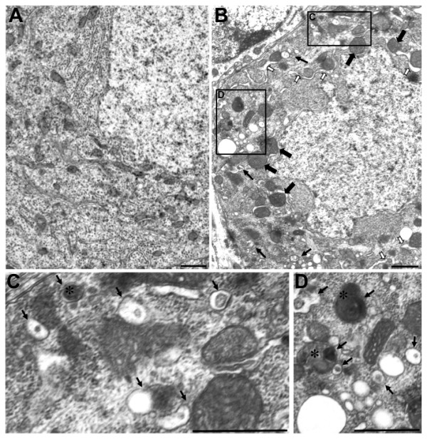
Ultrastructural analysis of pcd mice reveals altered mitochondrial morphology, endoplasmic reticulum histopathology, and autophagosome engulfment of mitochondria in Purkinje cell soma. (A-C) We performed an extensive ultrastructural comparison of Purkinje cell soma between 18 day-old wild-type and pcd5J homozygous mice. Representative micrographs are shown here. (A) Control micrographs from a wild-type Purkinje cell soma. (B) Purkinje cell somae in pcd5J homozygous develop huge mitochondria that are likely to represent fusion at early stages (filled black arrows) and very swollen endoplasmic reticulum that is largely denuded of ribosomes (white arrows). Autophagic vacuoles appear and engulf mitochondria and other organelles (line arrows). We noted a significant number of Purkinje cells from pcd5J homozygous mice undergoing dark cell degeneration which was accompanied by increased numbers of autophagic vacuoles. Dark cell degeneration was uncommon in wild-type control micrographs (not shown). (C) and (D) are higher magnification views of inset regions in (B). Autophagic vacuoles containing electron dense cytoplasmic elements (arrows) are present, including vacuoles containing mitochondria (asterisks). Scale bars represent 1 μM.