
Figure 4.
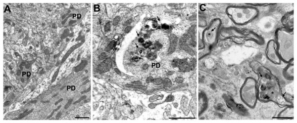
Ultrastructural analysis of pcd mice indicates accumulation of autophagic vacuoles and autophagosomes in Purkinje cell dendrites and axons. (A-B) We performed an extensive comparison of Purkinje cell dendrites between 18 day-old wild-type and pcd5J homozygous mice. Representative micrographs are shown here. (A) Dendrite process from a Purkinje cell neuron of a wild-type control. (B) In pcd5J homozygous mice, we noted frequent double-membrane bound structures in Purkinje cell dendrites (lined arrows), sometimes containing mitochondria (asterisks). (C) When we examined the Purkinje cell axons projecting to the deep cerebellar nuclei in pcd5J homozygous mice, we detected axons containing multiple autophagosome-like structures (arrows), containing mitochondria (asterisks) and other electron dense cellular components. Scale bars represent 1 μM.