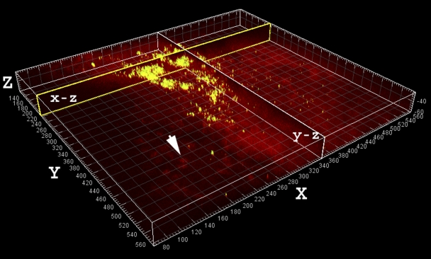
Fig. 2.
Large aggregate of viable (greenish-yellow) cocci in the aspirate stained with use of Molecular Probes LIVE/DEAD viability kit. The largest detected clumps were up to 100 μm in diameter and had a heterogeneous morphology consistent with that of “in vitro” grown Staphylococcus aureus biofilms. The aggregates may represent clumps of bacteria that had shed naturally from the biofilm, possibly contributing to systemic symptoms (e.g., fever). Sagittal sections through the clumps along the x and y axes are shown in the vertical planes, labeled “x-z” and “y-z.” The human nuclei (arrow) and associated tissue stained red. Scale: major divisions = 10 μm.

