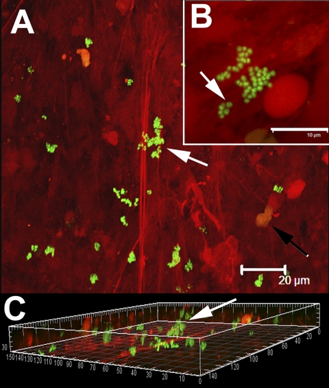
Fig. 3.
Single cells and clumps of viable cocci (stained green with use of Molecular Probes LIVE/DEAD viability kit) attached to tissue displaying characteristic staphylococcal morphology. Human tissue stained red with propidium iodide. A: Low-power image showing bacterial cells (arrow) attached to fibrous material (red striation). The nuclei of human cells were also visible (black arrow). Scale bar = 20 μm. B: Higher-power magnification of a group of cocci attached to fibrous material. Some cells were in the process of division (arrow), indicating that they were viable, which was consistent with the results of viability staining and culturing. Scale bar = 10 μm. C: Three-dimensional orthogonal projection of panel “A” showing that the biofilm clumps (arrow) were attached and protruding from the fibrous material. Scale: major divisions = 10 μm.

