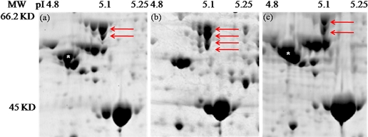
FIGURE 4.
Numerous highly abundant aphid and Buchnera proteins are differentially extracted by the TCA-acetone, phenol, and multi-detergent techniques using 2-DE. The images are taken from 24 cm, and 12% SDS-PAGE gels were stained with Colloidal blue and visualized using the Typhoon Variable Mode Imager Model 9400 (GE Healthcare). Numerous differences in protein spots and spot intensity are apparent. For example, red arrows point to various isoforms of the Buchnera protein, GroEL, in the (a) TCA-acetone extraction, (b) phenol extraction, and (c) multi-detergent extraction. White asterisks on the spots in the TCA-acetone (a) and multi-detergent (c) extraction indicate the highly abundant aphid protein β-tubulin, which is completely absent from the phenol extractions.

