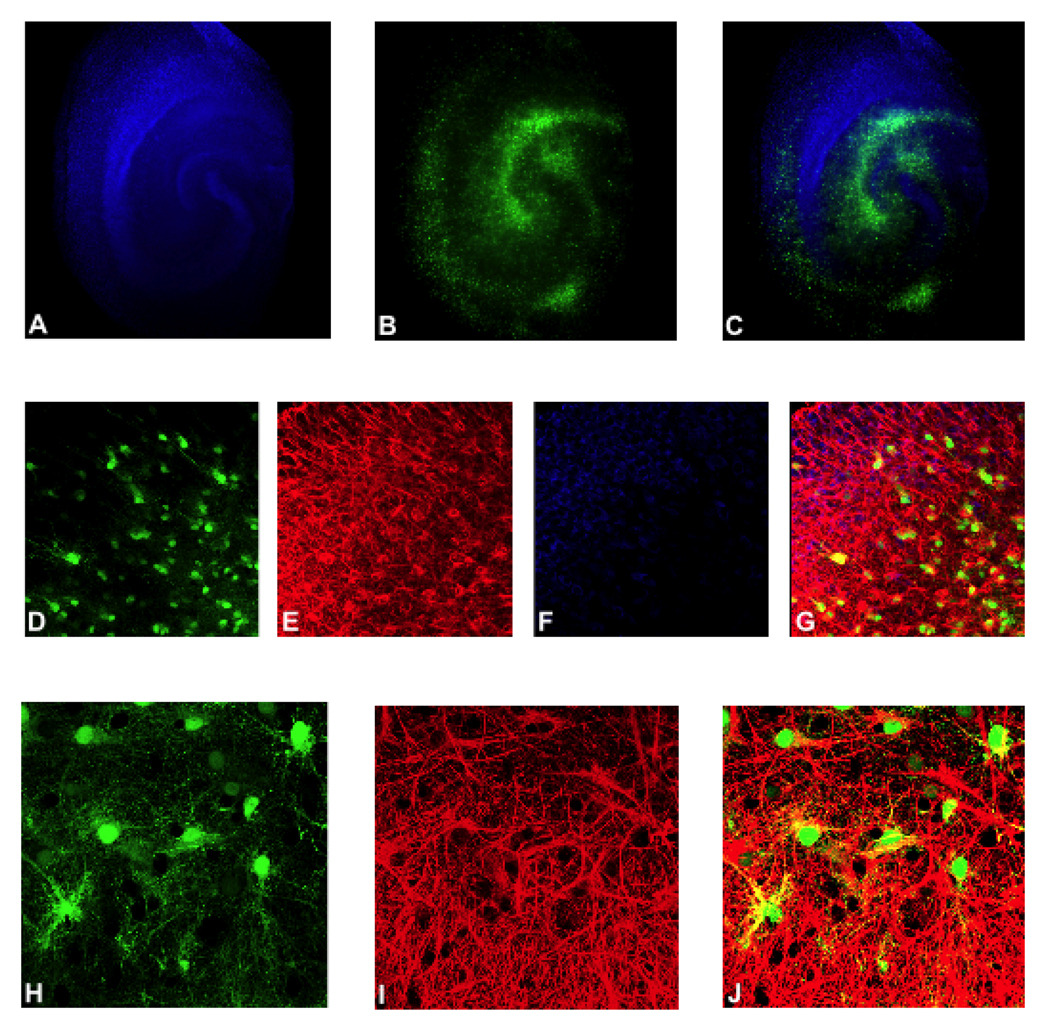
Figure 1. AAV1-GFAP construct targets transgene delivery and expression to astrocytes.
Representative images of rat hippocampal slice cultures (RHSC) transduced with AAV1-GFAP-GFP are shown. Panels A–C show low magnification images in which GFP expression (green) is observed throughout the slice culture and does not overlap with neurons stained with NeuroTrace (blue). Panels E, G, I and J show slice cultures stained with anti-GFAP antibody. Panels D–G are 40X images and panels H-J are 60X oil immersion images. Panels D,G, H and I show GFP positive astrocytes (green). Panels E, G, I and J show GFAP stained astrocytes and astrocytic processes (red); and panels G (merged image of panels D–F) and J (merged image of panels H–J) show colocalization of GFP and GFAP (yellow).

