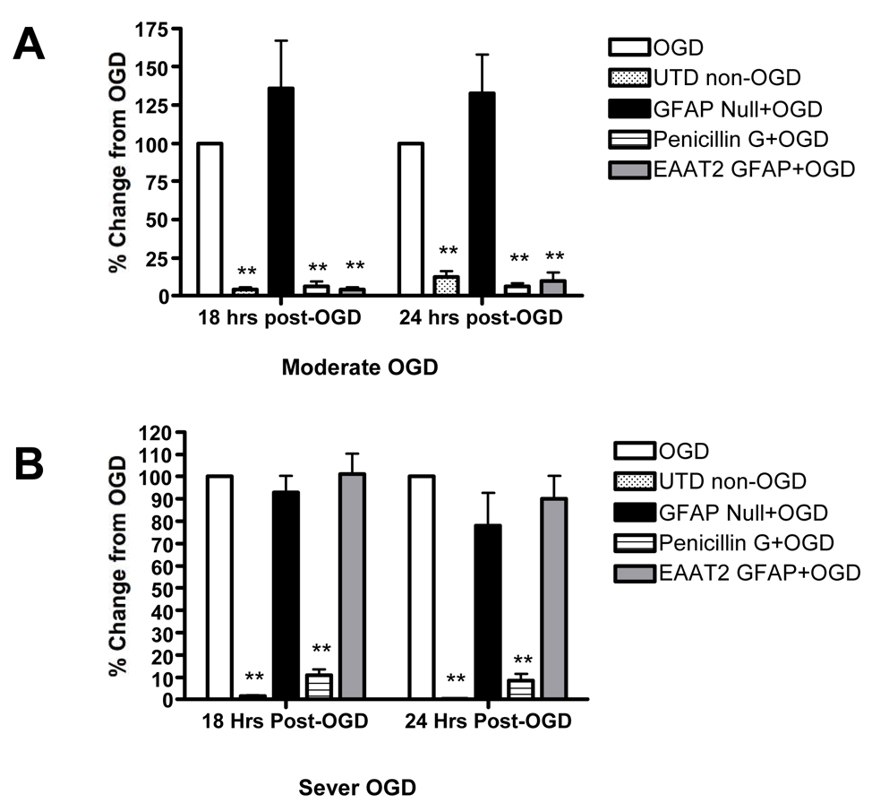
Figure 4. Over expression of exogenous EAAT2 in astrocytes enhances neuroprotection against oxygen glucose deprivation.
Neuronal damage mediated by OGD was measured by propidium iodide (PI) uptake in control RHSCs exposed to OGD (open white bars); RHSCs not exposed to OGD (stippled bars); RHScs transduced with AAV1-GFAP-null virus (solid black bars); RHSCs transduced with AAV1-GFAP-EAAT2 transduced (gray bars) and RHSCs treated with 100µM penicillin G (horizontal stripped bars). Panel A shows RHSCs exposed to moderate to OGD and imaged for PI fluorescence at 18, and 24 hours post OGD (n=10–13 slices). Panle B shows RHSCs exposed to sever insult and imaged for PI fluorescence at 18, and 24 hours post OGD (n=10–21 slices). (One-way ANOVA analysis, ** = p < 0.01)

