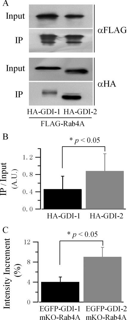
Figure 7. Rab4A favours binding to GDI-2.
(A) A representative result of the immunoprecipitation of GDIs with Rab4A as the bait protein. HEK-293 cells were co-transfected with FLAG–Rab4A(S27N) and HA–GDI-1 or HA–GDI-2. Immunoprecipitation (IP) and Western blotting were carried out in the same way as described in Figure 4(A). (B) Quantification of the interactions between GDIs and Rab4A. Results are represented as the means±S.E.M. of two independent experiments. Band intensity is normalized as described in Figure 4(B). (C) Quantification of the interaction of GDI-1 and GDI-2 with Rab4A by acceptor photobleaching FRET. HEK-293 cells were co-transfected with EGFP–GDI-1 or EGFP–GDI-2, and mKO–Rab4A(S27N). Experiments and data preparation were carried out in the same way as described in Figure 5(B). Results are represented as means±S.E.M. (GDI-1, n=25 cells; GDI-2, n=25 cells).

