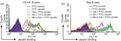
Figure 1.
Expression of Jacalin receptors on the surfaces of human primary CD19+ B cells and Raji B cells. CD19+ B cells from healthy human peripheral blood lymphocytes were separated with a QuadroMACS separation unit. The separated CD19+ B cells (a) and Raji B cells (b) were stained with fluorescein isothiocyanate-labelled Jacalin, and then analysed by flow cytometry, respectively. The cells were prepared and labelled as described in the Materials and methods. As a negative control, the autofluorescence of the cells was measured (purple area). As shown in each histogram, the sugar specificity of the binding of Jacalin to primary CD19+ B and Raji B cells was analysed in the presence of 50 mm Gal (pink), Glc (blue), or Me-α-Gal (orange), respectively.

