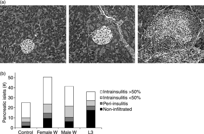
Figure 2.
(a) Representative examples of the classification of islets as non-infiltrated (left panel), peri-insulitis (middle panel) and intrainsulitis > 50% (right panel). (b) Mean total numbers of pancreatic islets counted from four slides. Pancreatic islets were classified as non-infiltrated, peri-insulitis, and intrainsulitis with less than or greater than 50% infiltrated lymphocytes. Mice were infected with either 40 L3 larvae (n= 6), implanted intraperitoneally with five adult female worms (n= 7) or five adult male worms (n= 5), or were sham-treated (n= 7). Combined data from two independent experiments are shown.

