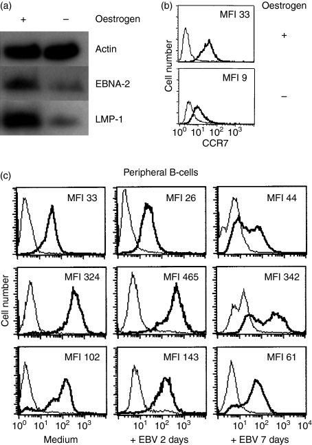
Figure 3.
Changes in CCR7 expression in ER/EB2-5 cells after oestrogen withdrawal. (a) CCR7 expression in proliferating ER/EB2-5 cells and ER/EB2-5 cells which have been cultivated in the absence of oestrogen for 3 days. The bold line represents the specific staining and the thin line the isotype-matched control. MFI = mean fluorescence intensity. (b) Western blot analysis of Epstein–Barr nuclear antigen 2 (EBNA2) and latent membrane protein 1 (LMP1). Actin was blotted as a loading control. Representative data of three independent experiments are shown. (c) CCR7 expression on peripheral B cells after in vitro infection with Epstein–Barr virus (EBV) for 2 and 7 days (n = 3). Approximately 75% of the cells were EBNA2 positive in the cultures on day 7. MFI = mean fluorescence intensity.

