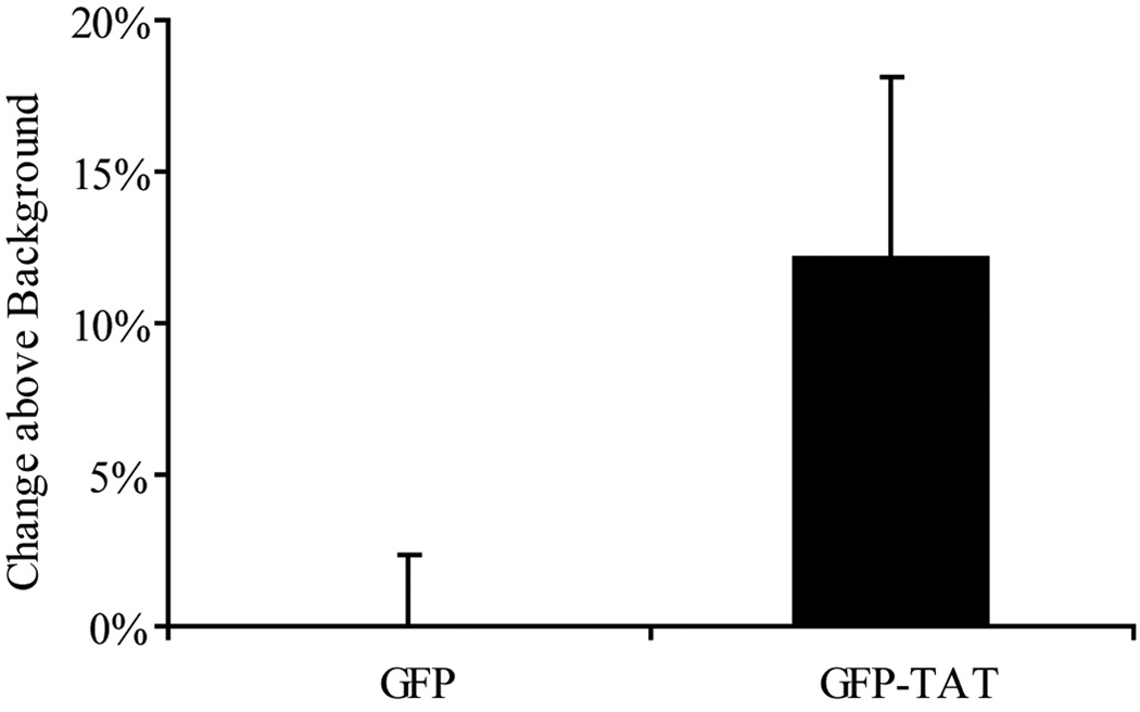
Fig. 4.
Transduction of GFP and GFP-TAT into neurons. Neurons in cocultures were incubated in 100 µg/mL GFP or GFP-TAT for 4 hours, and transduction was quantified using flow cytometry. Cell type was confirmed by Thy1 immunofluorescence after trypsinization. The percent increase in geometric mean fluorescence of Thy1+ cells compared to untreated control cells was calculated, to account for background autofluorescence (n>6, error bars: ±SEM). TAT did not significantly enhance GFP transduction into neurons, compared to GFP alone.

