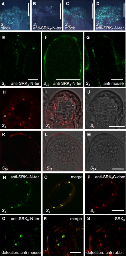
Figure 4.
Ligand-Induced Internalization of SRK3.
(A) to (D) Anti-SRK3-N-ter can functionally substitute for the natural SRK3 ligand in vivo. B. oleracea S3- and S29- stigmas were treated with anti-SRK3-N-ter or mock solution and pollinated with S15-pollen, which is normally compatible for both haplotypes. Pollen germination was visualized under UV light after aniline blue staining. Five independent experiments were performed, yielding similar results.
(A) Germination of S15-pollen on S3-stigma treated with mock solution. Bar = 500 μm.
(B) Rejection of S15-pollen on S3-stigma treated with anti-SRK3-N-ter solution. Bar = 500 μm.
(C) Germination of S15-pollen on S29-stigma treated with mock solution. Bar = 500 μm.
(D) Germination of S15-pollen on S29-stigma treated with anti-SRK3-N-ter solution. No rejection can be observed. Bar = 500 μm.
(E) and (F) B. oleracea S3- and S29-stigmas were treated with anti-SRK3-N-ter solution and pollinated with S15-pollen (normally compatible for both haplotypes). Antibody internalization was followed by immunofluorescence on fixed sections. The results could be reproduced in three independent experiments.
(E) Internalization in S3-papilla cells. Anti-SRK3-N-ter is found in intracellular compartments after 2.5 h, detected directly by the secondary anti-mouse antibody. Bar = 10 μm.
(F) No internalization is observed in S29-papilla cells. Bar = 10 μm.
(G) B. oleracea S3-stigmas were treated with secondary donkey anti-mouse Alexa555 solution and pollinated with S15-pollen. No internalization is observed in S29-papilla cells after 2.5 h. Bar = 10 μm.
(H) to (J) Detection of SRK3 on S3 stigma papilla sections. SRK3 is detected in intracellular compartments and unspecifically on the cell wall (arrow) by anti-SRK3C-dom antibody. Bright-field image is presented in (J) and the merge image in (I). Similar results were observed in three independent experiments. Bar = 10 μm.
(K) to (M) Absence of SRK3 from S29-stigma papilla sections (control experiment). The unspecific cell wall staining probably corresponds to the bands detected on protein gel blot with the same antibody. Bright-field image is presented in (M) and the merge image in (L). Similar results were observed in three independent experiments Bar = 10 μm.
(N) to (P) Simultaneous detection of SRK3 in S3-papilla cells using anti-SRK3-N-ter (recognizing the N terminus of SRK3) and anti-SRK3C-dom antibody (recognizing the C terminus of SRK3). Anti-SRK3C-dom does not recognize the PM-localized SRK3, possibly due to an interaction blocking the antibody epitope and shows a weak unspecific cell wall signal. Similar results were observed in three independent experiments. Bar = 10 μm.
(Q) to (S) Colocalization of internalized anti-SRK3-N-ter (detected directly by the secondary anti-mouse antibody) and SRK3 (detected by anti-SRK3C-dom and secondary anti rabbit antibody) in S3-papilla cells. Similar results were observed in three independent experiments. Bar = 10 μm.

