
Figure 2.
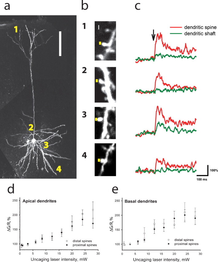
Glutamate uncaging reveals similar glutamate sensitivity of calcium transients in proximal and distal dendritic spines on thalamorecipient neurons in the ACx. a, The image of a thalamorecipient neuron filled with Fluo-5F and Alexa 594. Glutamate uncaging targeted dendritic spines in four different locations: (1) distal apical, (2) proximal apical, (3) proximal basal, and (4) distal basal. Scale bar, 100 μm. b, Representative images of dendritic spines in the four different locations. Yellow squares represent the sites of glutamate uncaging (0.5 ms exposure). Scale bar, 0.5 μm. c, Fluo-5F fluorescence intensity measured in dendritic spines (red) and parent dendritic shafts (green) from b in response to a single pulse of glutamate uncaging (arrow). d, e, Mean ± SEM peak Fluo-5F fluorescence (ΔG/R) intensity as a function of uncaging laser intensity measured in distal (open circles; n = 14 and 11) and proximal (filled circles; n = 12 and 17) dendritic spines of apical (d) and basal (e) dendrites, respectively.