
Figure 3.
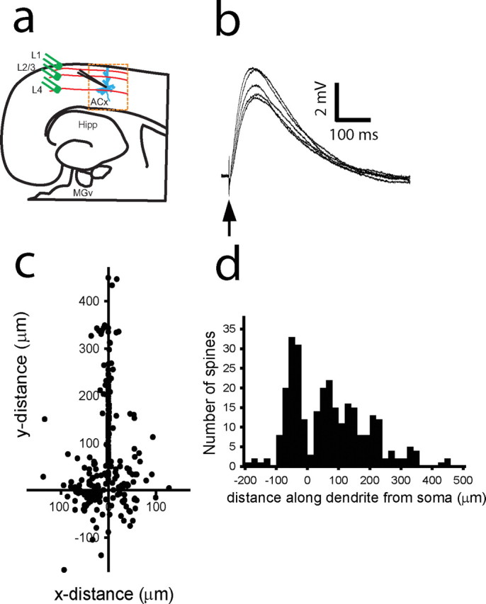
Map of active intracortical inputs on a dendritic tree of a thalamorecipient pyramidal neuron. a, Diagram of a thalamocortical slice containing parts of the MGv and ACx. Thalamorecipient pyramidal neurons (blue) are recorded in the ACx. Th afferents (red lines) are stimulated using stimulating electrodes (green). Hipp, Hippocampus. b, Monosynaptic EPSPs recorded in a thalamorecipient neuron in response to single cortical stimulation (arrow). c, Composite map of all active IC inputs (n = 272) detected from 87 thalamorecipient pyramidal neurons. Cartesian coordinates represent the sites of IC inputs on the dendritic tree, in which the coordinates of the soma are 0,0. Note that Cartesian coordinates of basal dendritic spines may be either in the positive or negative direction, depending on their orientation to the cell soma. d, Frequency histogram of the distance of all active IC inputs measured along the dendrites from the cell soma of thalamorecipient pyramidal neurons. Negative values denote basal dendrites, and positive values denote apical and oblique dendrites.