
Figure 4.
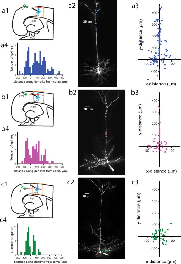
Maps of active intracortical inputs on dendritic trees of thalamorecipient pyramidal neurons found in response to L1, L2/3, or L4 stimulation. a1, b1, c1, Diagrams of a thalamocortical slice showing the site of L1 (a1), L2/3 (b1), and L4 (c1) stimulation relative to the recorded thalamorecipient neuron (blue). a2, b2, c2, Examples of Alexa 594 reconstructions of pyramidal thalamorecipient neurons showing locations (colored dots) in which active spines were detected in response to L1 (a2), L2/3 (b2), and L4 (c2) stimulation. a3, b3, c3, Cartesian coordinates represent the placement of L1-connected (a3; n = 145), L2/3-connected (b3; n = 83), and L4-connected (c3; n = 44) inputs along the dendritic trees, in which the coordinates of the soma are 0,0. a4, b4, c4, Frequency histograms of distances of active L1 (a4), L2/3 (b4), and L4 (c4) inputs measured along dendrites from the cell soma of thalamorecipient pyramidal neurons. Negative values denote basal dendrites, and positive values denote apical and oblique dendrites. Hipp, Hippocampus.