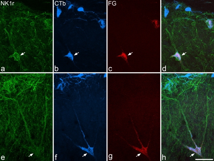
Figure 9.
Retrograde labelling of lamina III/IV NK1r-immunoreactive neurons. These images are from confocal scans that show immunoreactivity for NK1r (green), CTb (transported from LPb, blue), and Fluorogold (FG; transported from thalamus, red) in transverse sections through C7 (a–d) and L4 (e–h) from experiment Pb3. In each case, a single large NK1r-immunoreactive cell with its soma in lamina III or IV is labelled with both retrograde tracers (arrows). Images are projections of 20 (a–d) or 18 (e–h) confocal optical sections at 2-μm z-spacing. Scale bar = 50 μm.

