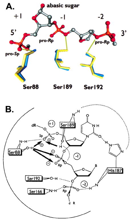
Figure 21.
Direct and indirect coupling energies between serine side chains and phosphodiester oxygens of UDG. (A) The interactions of UDG with its abasic site DNA product as determined by X-ray crystallography (pdb code 1FLZ, yellow; pdb code 1SSP, blue). The coupling energy may be interpreted as the additional apparent energy of interaction between the phosphodiester oxygen and the given serine residue as compared to that of the interaction with sulfur (Table 1). In this figure, bold arrows with plus and minus signs indicate cooperative and anticooperative coupling energies, respectively. Dashed lines represent hydrogen bonds. The S88A mutation causes an increased Rp and Sp thio effect at the −1 position (strain), and the S189A mutation causes a stereoselective decrease in the Sp thio effect at the +1 phosphodiester (positive cooperativity). The S189A and S192G mutations have little effect on the magnitude of the thio effects at the −1 or −2 position. A highly cooperative network is indicated.

