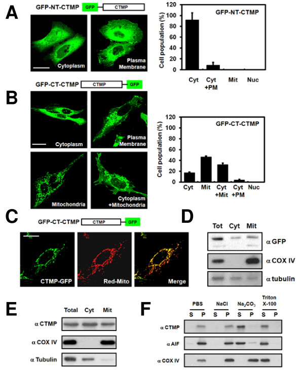
Figure 2.
Functional mitochondrial localization of CTMP. U2OS cells were transfected with (A) CTMP GFP-tagged at the N-terminus (GFP-NT-CTMP) or (B) CTMP GFP-tagged at the C-terminus (GFP-CT-CTMP) for 24 h. Differential localization of CTMP (Cyt: cytoplasm, PM: plasma membrane, Mit: mitochondria, Nuc: Nucleus) was examined using confocal microscopy. At least 200 cells were counted from three distinct fields for each transfected group. U2OS cells were co-transfected with (C) pEGFP-N3-CTMP and pDsRed-mito for 24 h and analyzed by confocal microscopy. (D) U2OS cells were transfected with pEGFP-N3-CTMP. Total lysates (Tot), cytosolic fractions (Cyt), and mitochondrial fractions (Mit) were analyzed by immunoblot analysis. (E) The subcellular fractions of HEK 293 cells were analyzed with the indicated antibodies. (COX IV, Mit marker; α-tubulin, Cyt marker). (F) Mitochondrial fractions of HEK 293 cells were isolated and treated under the indicated conditions. Samples were separated into the supernatant (S) and precipitate (P) fractions and then analyzed. (AIF, Mit intermembrane space marker).

