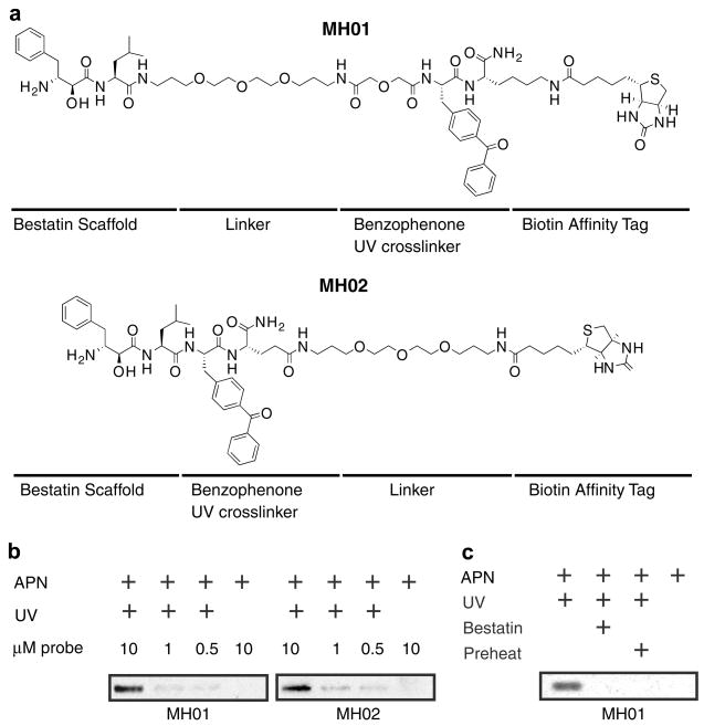
Figure 2.
Anatomy of ABPs and labeling of aminopeptidase N. (a) Structure of ABPs MH01 and MH02. (b) Aminopeptidase N (0.13 U) was treated with 10, 1, or 0.5 μM of either MH01 or MH02 for 1 h in 50 mM Tris–HCl, pH 7.8, 0.5 μM ZnCl2 (buffer A). Certain reaction mixtures were UV crosslinked for 1 h on ice. Reactions were quenched with SDS–PAGE buffer, and labeled protein was visualized via SDS–PAGE and western blotting for biotin. (c) Aminopeptidase N was treated with 100 μM of the aminopeptidase inhibitor bestatin or DMSO for 1 h in buffer A followed by labeling with MH01 for 1 h. Reactions were UV crosslinked (or not) for 1 h on ice, and labeled protein was visualized as in Figure 2b.

