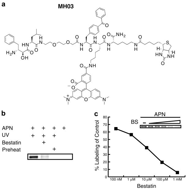
Figure 3.
Aminopeptidase N ABP labeling by fluorophore-containing bestatin-based ABP. (a) Structure of MH03 probe. (b) Labeling of aminopeptidase N was performed as described in Figure 2, but using MH03 and visualized using SDS–PAGE and in-gel fluorescent scanning. (c) Aminopeptidase N was pretreated with multiple concentrations of bestatin (BS) for 1 h and then labeled with 10 μM fluorescent MH03. After in-gel fluorescent scanning labeling was quantified using ImageQuant software (GE).

