
Figure 4.
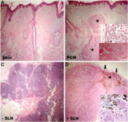
Histology of FFPE specimens evaluated for their small RNA profile. Samples were derived from a 52-year-old man with PCM, 4.0 mm and level IV, on the scalp and his sentinel nodes. A: Uninvolved skin >2.0 cm away from the primary tumor in a wide local excision. B: Residual primary melanoma (asterisks) in the wide local excision. The upper inset shows a high magnification of tumor cells and the lower inset shows a segment of skeletal muscle, identified on the slide, correlating with the expression of miR-1. C: SLN negative for metastasis. D: SLN positive for metastasis. A rim of nodal tissue remains (arrows) as the large metastatic tumor replaced the nodal architecture. A large focus of melanin pigment was identified (asterisk). The inset shows a high magnification of tumor cells. Original magnification: A–D, ×40; insets in B and D, ×400.