
Figure 6.
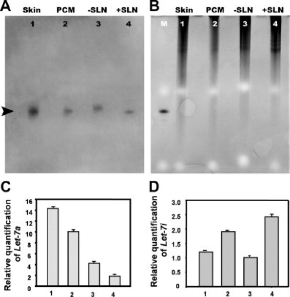
miRNAs were well-preserved in 10-year-old formalin-fixed FFPE specimens. A: Northern blot analysis shows the expression of the human let-7a in all samples from a 52-year-old man with scalp melanoma, corresponding to the histology shown in Figure 4: skin, PCM, −SLN, and +SLN. Each sample (4 μg/lane) was loaded and resolved on a denaturing 15% polyacrylamide gel. A 21/22-bp band is identified (arrowhead) corresponding to the size of lin-4 in B. B: For loading control, the 15% polyacrylamide gel for Northern blot was stained by EtBr before transferring to membrane. Lane M lane represents synthetic Caenorhabditis elegans miRNA, lin-4 loaded for size comparison. C: Quantitative real-time RT-PCR demonstrated down-regulation of let-7a expression in PCM and +SLN, when compared with normal skin and −SLN. D: The opposite effect, up-regulation, was demonstrated for let-7i expression in PCM and +SLN, when compared with normal skin and −SLN. The amount of miRNA was normalized using U6. Each reaction was performed in triplicate; error bars show standard deviations.