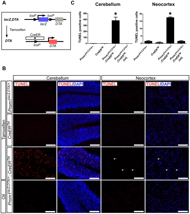
Figure 3. Tamoxifen-dependent Cre activation induces apoptosis of Prom1-expressing cells in vivo.
(A) Schematic diagram of DTA activation. Tamoxifen-dependent Cre activation deleted the lacZ gene cassette, leading to the induction of DTA expression upon activation of the Prom1 promoter. (B) Intraperitoneal injection of tamoxifen-induced cell death (red) in cerebellum (left panels) and neocortex in telencephalon (right panels) as well as midbrain and white matter (not shown). Arrow heads indicate TUNEL-positive cells in the neocortex. All nuclei were counterstained with DAPI (blue). Scales, 100 µm. (C) The number of TUNEL-positive cells per section were counted in the whole neocortex and in half of one lobule of the cerebellum. The results shown are the mean±s.e.m. of two independent experiments. *P<0.05. Similar results were also observed in the midbrain and white matter (not shown).

