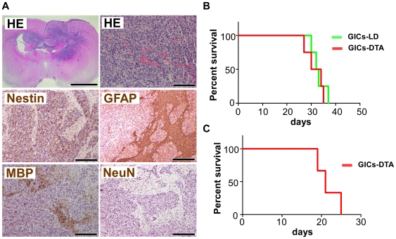
Figure 7. Prom1-expressing cells are not required for the tumorigenesis of GICs in nude mice.
(A) Brain sections with tumors derived from GICs-DTA. The GICs invaded into the brain parenchyma. HE staining of the tumors shows necrosis, hypercellularity and hypervascularity, multinuclear giant cells, and mitotic cells; these pathological features are similar to human GBM (upper panels). Immunohistochemical analysis of the tumor for Nestin, GFAP, MBP, and NeuN (middle and lower panels). Scales, 2 mm (upper left panel), 100 µm (upper right panel), and 200 µm (middle and lower panels). (B) Survival curves for mice (n = 4 of each line) injected with 104 GICs-LD (green line) and GICs-DTA (red line). No significance was observed, P = 0.65. (C) Survival curve for mice (n = 3) serially transplanted with 104 GFP-positive primary tumor cells.

