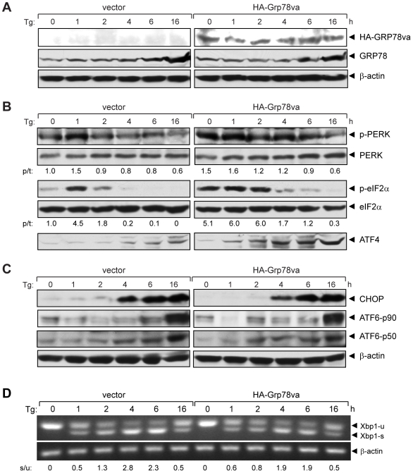
Figure 4. GRP78va enhances PERK signaling.
A. Western blots to confirm the ectopical expression of GRP78va. HeLa cells stably transfected with vector or pcDNA/HA-Grp78va were treated with Tg (300 nM) for the indicated time. HA-GRP78va and endogenous GRP78 were detected by anti-HA and anti-GRP78 antibody respectively. β-actin levels served as loading control. B. Western blots were performed to detect the kinetics and magnitude of UPR signaling markers identified on the right following Tg treatment for the indicated time points. The experiments were repeated twice. The representative results are shown with the ratio (p/t) of p-PERK/PERK or p-eIF2α/eIF2α indicated under each panel with the non-treated control cells set as 1.0. C. Detection of the CHOP activation and ATF6 cleavage following Tg treatment for the indicated time points. The uncleaved (p90) and cleaved (p50) forms of ATF6 are indicated. β-actin levels served as loading control. D. RT-PCR to detect Xbp1 splicing following Tg treatment for the indicated time. The positions of the Xbp1 unspliced (u) and spliced (s) forms were indicated and the ratio of spliced to unspliced form (s/u) is indicated below. The experiments were repeated three times and the representative results are shown.

