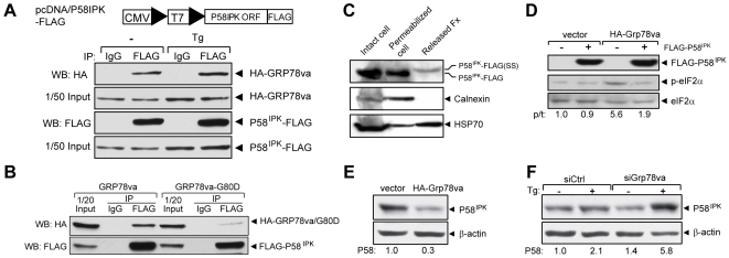
Figure 6. Physical and functional interactions between GRP78va and P58IPK.
A. Co-immunoprecipitation of GRP78va with P58IPK. 293T cells co-transfected with pcDNA/P58IPK-FLAG (schematic drawing shown above) and pcDNA/HA-Grp78va were either non-treated (−) or treated with Tg (300 nM) for 16 hours. Immunoprecipitations were performed with either anti-FLAG antibody or control mouse IgG, followed by Western blot with the respective antibodies. The input levels are shown. B. Reduced binding between FLAG-P58IPK and ATP binding mutant of GRP78va. 293T cells were co-transfected with pcDNA/FLAG-P58IPK and pcDNA/HA-Grp78va or the mutant Grp78va-G80D. Immunoprecipitations were performed with anti-FLAG antibody or control IgG, followed by Western blot. C. Cytosolic localization of P58IPK determined by the cell permeabilization assay. HeLa cells transiently transfected with pcDNA/P58IPK-FLAG were permeabilized by 0.01% digitonin for 5 minutes. The various fractions were subjected to Western blots for the proteins as indicated. P58IPK-FLAG(SS) designates P58IPK with a slower electrophoretic mobility being released from the permeabilized cells, consistent with retention of signal sequence. D. Overexpression of FLAG-P58IPK suppressed eIF2α phosphorylation mediated by GRP78va. Following transient transfection of the P58IPK expression plasmid or vector into the indicated stably transfected HeLa cell lines, Western blots were performed to probe for the indicated proteins. The experiments were repeated twice. The representative Western blots are shown with the ratio (p/t) of phospho-eIF2α to total eIF2α indicated. E. Overexpression of GRP78va reduced endogenous P58IPK levels. Western blots were performed in HeLa cells stably transfected with vector or HA-GRP78va expression plasmid. The experiments were repeated three times. The representative Western blots are shown and the P58IPK levels normalized to β-actin are indicated below. F. Knockdown of GRP78va increased endogenous P58IPK level. Western blots were performed with HeLa cells transfected with control siRNA (siCtrl) or Grp78va siRNA (siGrp78va) to detect endogenous P58IPK and β-actin level. The cells were either non-treated (−) or treated (+) with Tg (300 nM) for 16 hours. The P58IPK levels normalized to β-actin are shown below the immunoblots. Statistical comparisons were made between Tg-treated cells transfected with siCtrl or siGrp78va, p = 0.03.

