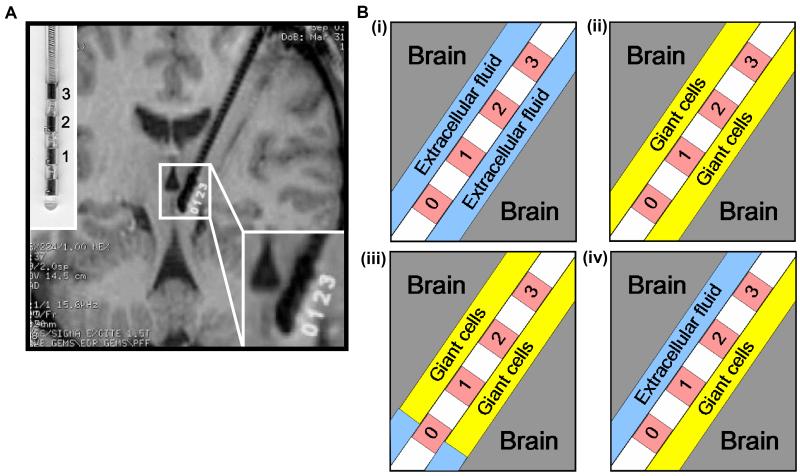
Figure 1.
Structural definition of the electrode-brain interface (EBI): (A) an MRI of the implanted quadripolar DBS electrode in situ, with an enlarged photo of the electrode model 3387 (the left upper corner) and an enlargement of the electrode-brain interface (the right lower corner), and (B) schematic representation of the EBI consisting of three essential elements of the implanted quadripolar electrode (contact 0 to 3, with contact 0 activated), the surrounding neural tissue, and the “peri-electrode space” in between, which is filled with ECF in the acute stage (i), with giant cells in the chronic stage (ii), and a mixture of ECF and giant cells in the transitional stages post-implantation. The giant cells grow transversely over all but the tip of the electrode (iii), or the giant cells grow longitudinally over the lower half of the electrode (iv).

