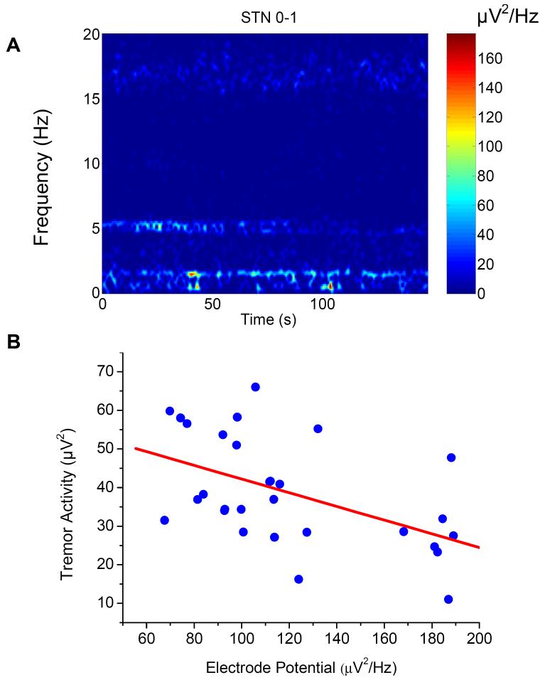
Figure 3.
A representative bipolar (contact 0-1) LFP recording from the subthalamic nucleus (STN) of a patient with Parkinsonian tremor. A: Three frequency components of the modulated electrode potential (1.0 - 1.5Hz), tremor oscillation (4 to 6Hz) and the beta oscillation (15 to 20Hz) in the time-frequency spectrograms of compound LFPs; and significant reverse correlations in power density between the electrode potential and the tremor oscillation (B) but not the beta oscillation.

