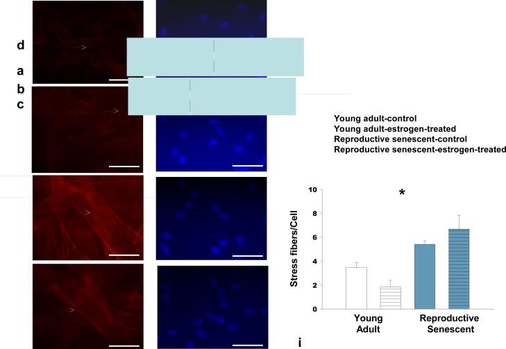
FIG 2.
F-actin stress fibers are significantly increased in reproductive senescent astrocytes as compared to young adult astrocytes. Stress fiber formation was analyzed in astrocytes cultured in the presence and absence of 40 nM estrogen by Alexa Fluor 594® phalloidin staining in young (a,b,i) and reproductive senescent astrocytes (c,d,i) and counter-stained with the nuclear dye Hoechst 33258 (e-h). Estrogen treatment attenuated stress fiber formation in young adults but not reproductive senescent astrocyte cultures. White arrow indicates presence of stress fibers. Bars represent mean ± SEM of a single representative experiment with n=4-5 for each treatment group. Interaction of age and hormone is statistically significant at p<0.05 and indicated by (*). Bar: 50 μm.

