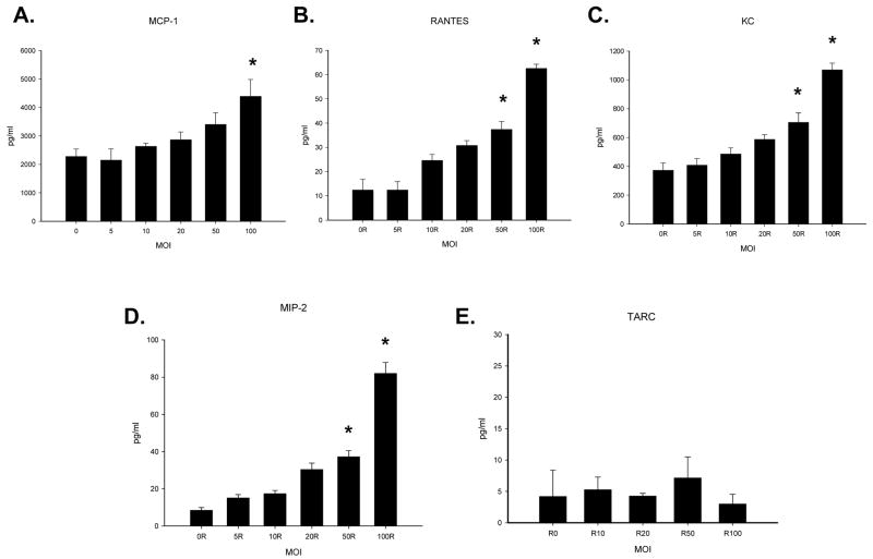
Figure 2. Chemokine levels in the supernatant (panels A–E) of in vitro cholangiocytes infected with increasing amounts of RRV.
In vitro, cholangiocyte cells were infected with increase amounts of RRV (n=3–5 tubes). Chemokine expression in the supernatants was determined by ELISA and expressed as means with standard error. Some chemokine expression was seen in the absence of infection in controls, though virus at MOIs of 50 and 100 resulted in significant increases in the quantity of MIP-2, RANTES and KC found in supernatant fluid from cholangiocytes (p<0.05) 24 hours after infection. MCP-1 was found to be significantly elevated over background expression at an MOI of 100 (p<0.05). Experiment conditions repeated 3 times.

