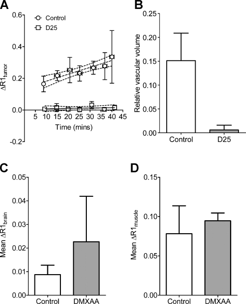
Figure 3.
Reduction in vascular volume of orthotopic HNC xenografts after VDA treatment. (A) Temporal change in R1 (ΔR1) values of control and DMXAA-treated orthotopic FaDu tumors calculated during a 40-minute period showing a significant decrease in contrast agent accumulation between control and treatment groups (P < .001 between slopes). (B) Calculated RVV of untreated control tumors and DMXAA-treated tumors showed a marked reduction 24 hours after DMXAA treatment compared with untreated controls (P < .05), indicative of significant tumor vascular disruption by DMXAA. No significant differences were seen (P > .5) in the calculated ΔR1 values of murine brain (C) and muscle tissues (D) from animals in the control and treatment groups.

