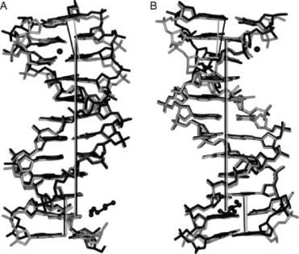
Figure 4.

Superimposition of the DDD (PDB entry BDL084) (black) and DDDZ10 (gray) duplexes. The view is into the (A) minor groove and (B) major groove, with respect to the central base pairs. Mg2+ and a partial spermine molecule in the structure of the native DDD are shown in a ball-and-stick representation. The helix axes are based on the central hexamer duplexes and are coincident (straight gray and black lines for DDDZ10 and DDD, respectively). In both duplexes, the helix axes defined by base pairs G10 · C15, C11 · G14, and G12 · C13 at one end are parallel with those of the central hexamers. Conversely, the helix axis defined by base pairs C1 · G24, G2 · C23, and C3 · G22 at the other end is inclined more in the native DDD than in DDDZ10 relative to the respective helix axis.
