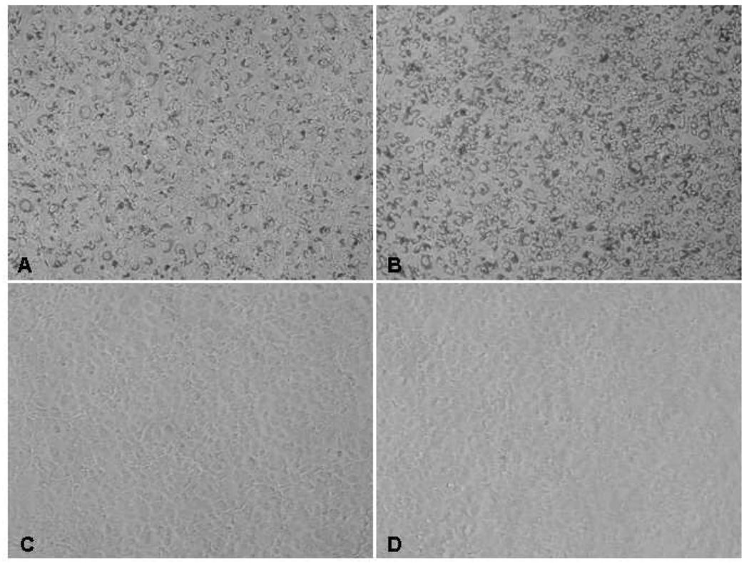
Figure 2. Phase-contrast microscopy images of human corneal endothelial cells (HCECs) in monolayer culture with and without incorporated superparamagnetic microspheres (SPMs).
After overnight incubation, confluent HCECs in monolayer were incorporated with A) 900 nm SPMs at 500 SPMs per cell plated, B) 900 nm SPMs at 500 SPMs per cell plated, and C) 100 nm SPMs at 16 µL per culture well. The 900 nm SPM was easily visible in the cells, whereas the 100 nm SPM was not, with the latter appearing similar to HCECs without SPMs (D). At confluence, HCECs assume a near hexagonal morphology similar to that of corneal endothelium in vivo. The cells shown are from one donor in passage 2; magnification, 100x.

