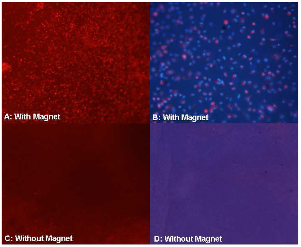
Figure 7. Fluorescence microscopy of posterior corneal stroma after human corneal endothelial cell (HCEC) transplantation.
A. Many DiI-labeled (red) donor HCECs were detected on corneas of anterior segments subjected to the magnetic field (magnification, 40x). B. At higher magnification (200x), donor HCEC density was 981 cells/mm2 in this recipient (nuclei are stained blue with DAPI and donor cell cytoplasm is stained red with Di-I). C and D. No donor HCECs were detected on control corneas not subjected to a magnetic field (magnification, 40x and 200x, respectively).

