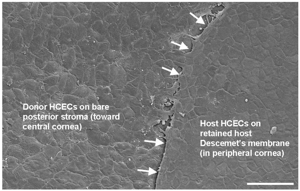
Figure 9. Formation of a confluent monolayer of human corneal endothelial cells (HCECs) on recipient human corneal stroma.
Scanning electron microscopy showed donor HCECs flattening and establishing a confluent monolayer on bare corneal stroma 3 days after transplantation; donor HCEC density was 1,525 cells/mm2. Donor HCECs were incorporated with 100 nm superparamagnetic microspheres and were transferred to the recipient human anterior segment in the presence of a magnetic field for 48 hours. Host HCECs on retained Descemet’s membrane were present peripherally; the stripped edge of Descemet’s membrane was evident (arrows). Bar, 100 µm.

