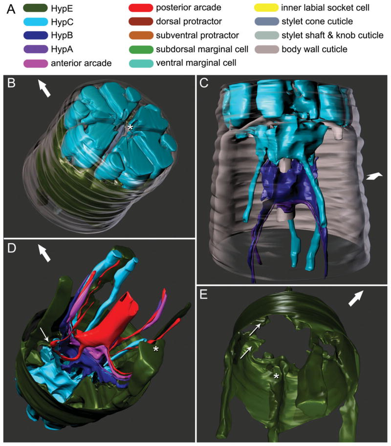
Fig. 2.
Three-dimensional reconstructions based on serial TEM sections of tissues and cuticle of the stomatostylet apparatus of Aphelenchus avenae. Body wall cuticle and all somal extensions are truncated posteriorly; body wall cuticle and most anterior epidermis (i.e., HypC) are truncated anteriorly through the tips of the lips. Large arrows indicate dorsal. A: Key to cells (applies also to Fig. 3). B: Oblique anterior view of HypE toroid, HypC toroid, and the tip of the stylet, all seen through the body wall cuticle (rendered transparent). Asterisk indicates stomatal opening. C: Ventral view of HypC, HypB, and HypA toroids lining the cephalic framework (rendered transparent) and the vestibule extension. D: Oblique posterior view of all anterior syncytia, including extensions in pseudocoelomic cords. Thin arrow indicates point of contact between lateral somal extension of HypE and ventrolateral somal extension of the posterior arcade syncytium; asterisk indicates incipient groove in somal extension of HypE that envelops ventral pseudocoelomic cord posteriorly. E: Oblique posterior view of HypE toroid, showing some cords and other medial structures. Thin arrows indicate examples of pockets for attachment of somatic and stylet protractor muscles; asterisk indicates ventral loop through which protractors meet.

